

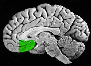
Medial Prefrontal Cortex
 |
|
PI Name: | BOTTERON, KELLY N. |
|
MALLIKRODT INSTITUTE OF RADIOLOGY |
Project Title: | NEUROMORPHOMETRY IN DEPRESSION |
|
|
|
|
Abstract:
My research investigates structural brain differences in children with affective disorders and attention deficit disorder. Populations of interest include major depression and attention deficit hyperactivity disorder in children and adolescents. Currently, we are examining structural MRI differences in discordant twin populations. In addition, in order to better understand structural abnormalities which we and others are demonstrating in children with psychiatric disorders, we are seeking to better characterize the progress of normal structural development, by MRI, in normal control populations including twins of children and adolescents.
Secondary to ongoing neurodevelopmental changes, image analysis methodologies need specific validation and potential modifications for use in child populations. We are involved with Dr. Michael Miller's lab (Johns Hopkins University) on the application of newer image analysis techniques, including automated 3-D, high resolution, warping atlases for child and adolescent populations.
Download:
This data is available with registered usernames and passwords. If you have already registered, and your registration has been confirmed, you may proceed to the download page.
References:
Miller MI, Massie A, Ratnanather, Botteron KN, and Csernansky JC: Bayesian construction of geometrically based cortical thickness metrics. NeuroImage. 12:676-687, 2000.
Ratnanather JT, Botteron KN, Nishino T, Massie AB, Lal RM, Patel SG, Peddi S, Todd RD, and Miller MI: Validating Cortical Surface Analysis of Medial Prefrontal Cortex metrics. NeuroImage. 14:1058-1069, 2001.
Brain Subvolume Image Data for Validation of Segmentation:
5 subvolumes are available in two formats: ANALYZE (7.5) and MATLAB formats.
The MATLAB data was generated on Linux but can be read in by other *IX systems.
Explanation of MATLAB format:
- mrinz - nonzero mri intensity voxel
- mannz - corresponding hand segmented voxel
- coord - voxel coordinate identifier
- ic - x coordinate: if ic value is zero set to xdim
- jc - y coordinate: if jc value is zero set to ydim
- kc - z coordinate: if kc value is zero set to zdim
- xdim, ydim, zdim - voxel resolution
Hand segmentation labels:
- CSF = 1
- CSF/GMA = 2 (partial volume with equal probability)
- GMA = 3
- WMA = 4
Supported by:
- NIH R01-MH062626
- NIH P41-RR15241
- NSF NPACI
For additional information or questions, please contact cis@cis.jhu.edu