Cortical Modeling
Describing the surface of the human brain is a complex task in that no two brains are identical and so there is no latitude-longitude equivalent in comparing structures between two brains.
Thus, in order to see how a diseased brain differs from a normal brain, or in comparing two normal brains, a common coordinate system must exist so that one map can be compared to another. Hence the exploding area of cortical cartography.
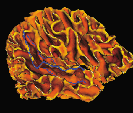
Dynamic programming tracking of the superior temporal gyrus in a cortical surface of brain from Laboratory of NeuroImaging (LONI) at UCLA (displayed here) shows where the points of maximal and minimal curvature (gyri and sulci respectively) occur.
As the curvature of the cortex becomes increasingly negative, the visual region presented becomes increasingly dark. Therefore, the darker the region, the greater the negative curvature.
Dynamic programming methods were used to define the locus of points of maximum or minimum curvature of the temporal love shown here and on the previous page.
